Optical Microscopy Application: Darkfield Illumination
저자: Stephan Briggs
Darkfield illumination is a technique in optical microscopy that eliminates scattered light from the sample image. This yields an image with a dark background around the specimen, and is essentially the complete opposite of the brightfield illumination technique. The primary imaging goal of the darkfield illumination technique is to enhance the contrast of an unstained sample, which is incredibly powerful, yet simple, for live cellular analysis or samples that have not gone through the staining process.
Image Appearance
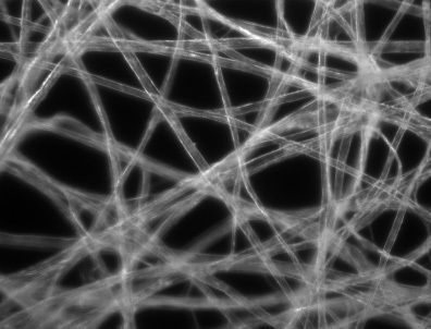
A typical darkfield illumination image has a white/bright specimen with a dark background and environment filling the image. This is the exact opposite of a brightfield illumination image, and is useful for unstained specimens or images that require increased contrast. The advantage with using darkfield illumination is that unstained specimens can remain alive and vital, whereas their brightfield counterparts must be treated and are no longer active. Also, it is possible to acquire more qualitative results with this technique through live cellular analysis. For additional information on the brightfield technique, please read Optical Microscopy Application: Brightfield Illumination.

Figure 1: Darkfield Illumination Image of Tissue Paper

Figure 2: Optical Path for Darkfield Illumination
Technical Details
The light path of the darkfield illumination technique is typically applied to an upright microscope, as seen in Figure 2. The light path consists of three key components.
- Light Source: enters the microscope and hits the dark field patch stop, which is a disc used to block light from entering the condenser and leaves a circular ring of illumination.
- Condenser Lens: collects outer ring of illumination and focuses it on the sample.
- Objective Lens: light hits the sample, and is transmitted or scattered from it. Scattered light enters the objective lens, whereas transmitted light is not collected by the lens. The direct illumination block assists in this step.
Additional optical microscopy applications include brightfield illumination, phase contrast, fluorescence, and differential interference contrast.


본사 및 지사별 연락처 확인하기
견적 요청 도구
재고 번호 입력 필요
Copyright 2023, 에드몬드옵틱스코리아 사업자 등록번호: 110-81-74657 | 대표이사: 앙텍하우 | 통신판매업 신고번호: 제 2022-서울마포-0965호, 서울특별시 마포구 월드컵북로 21, 7층 (서교동, 풍성빌딩)